ARTHRITIS IN CATTLE
373
It.:-:_.
~,t
e_ .,.-
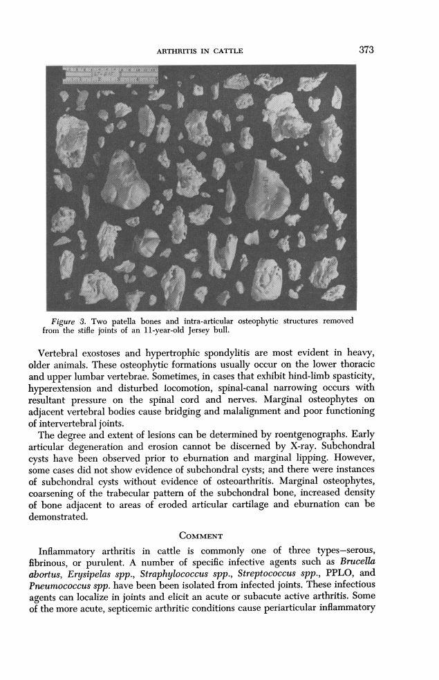
Figure 3. Two patella bones and intra-articular osteophytic structures removed
from the stifle joints of an 11-year-old Jersey bull.
Vertebral exostoses and hypertrophic spondylitis are most evident in heavy,
older animals. These osteophytic formations usually occur on the lower thoracic
and upper lumbar vertebrae. Sometimes, in cases that exhibit hind-limb spasticity,
hyperextension and disturbed locomotion, spinal-canal narrowing occurs with
resultant pressure on the spinal cord and nerves. Marginal osteophytes on
adjacent vertebral bodies cause bridging and malalignment and poor functioning
of intervertebral joints.
The degree and extent of lesions can be determined by roentgenographs. Early
articular degeneration and erosion cannot be discerned by X-ray. Subchondral
cysts have been observed prior to eburnation and marginal lipping. However,
some cases did not show evidence of subchondral cysts; and there were instances
of subchondral cysts without evidence of osteoarthritis. Marginal osteophytes,
coarsening of the trabecular pattern of the subchondral bone, increased density
of bone adjacent to areas of eroded articular cartilage and eburnation can be
demonstrated.
COMMENT
Inflammatory arthritis in cattle is commonly one of three types-serous,
fibrinous, or purulent. A number of specific infective agents such as Brucella
abortus, Erysipelas spp., Straphylococcus spp., Streptococcus spp., PPLO, and
Pneumococcus spp. have been been isolated from infected joints. These infectious
agents can localize in joints and elicit an acute or subacute active arthritis. Some
of the more acute, septicemic arthritic conditions cause periarticular inflammatory